The complete radiology platform for veterinarians
Built with veterinarians and developed alongside leading equine clinics. Transform your diagnostic workflow with AI-powered radiology tools.
From X-ray to report, in one complete workflow
Capture, analyze, and document radiology findings with AI-powered precision. Streamline your diagnostic process from image acquisition to final report.
Capture
Upload X-ray images directly from your device
Quality Check
AI validates image diagnostic quality
Anomaly Detection
AI identifies potential abnormalities
Measurements
Automated anatomic measurements
Report
Generate structured reports
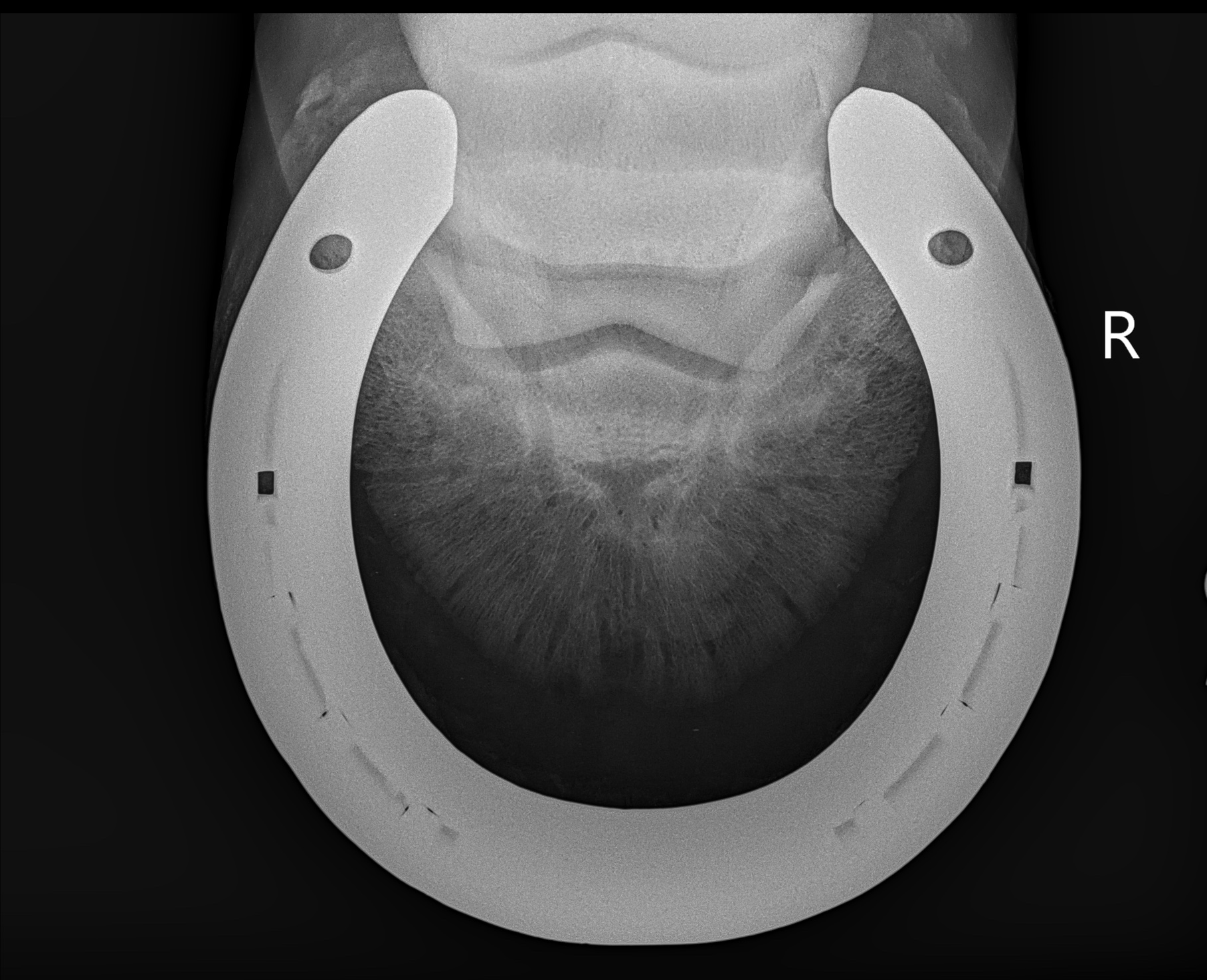
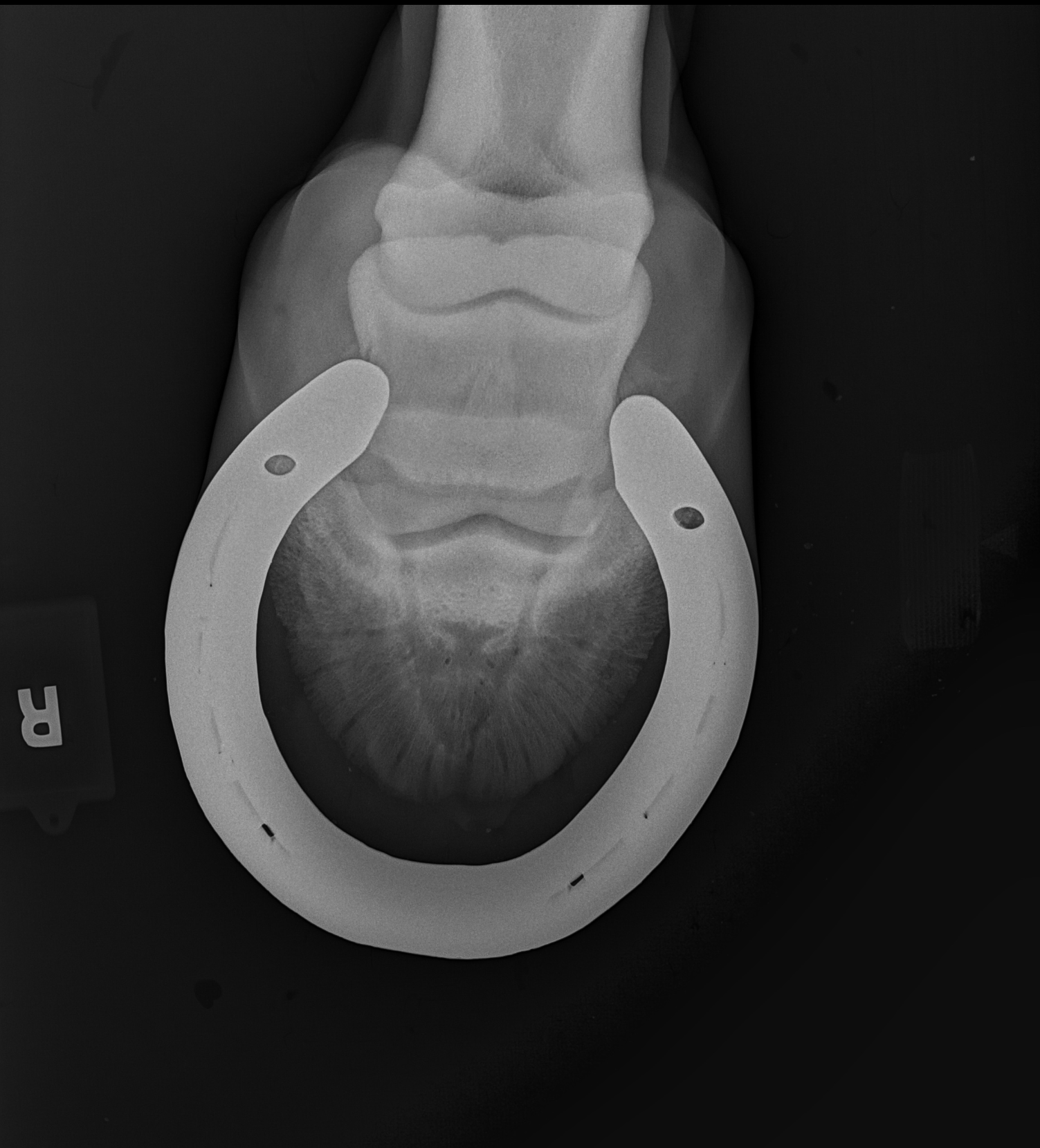
Compare
Track changes over time
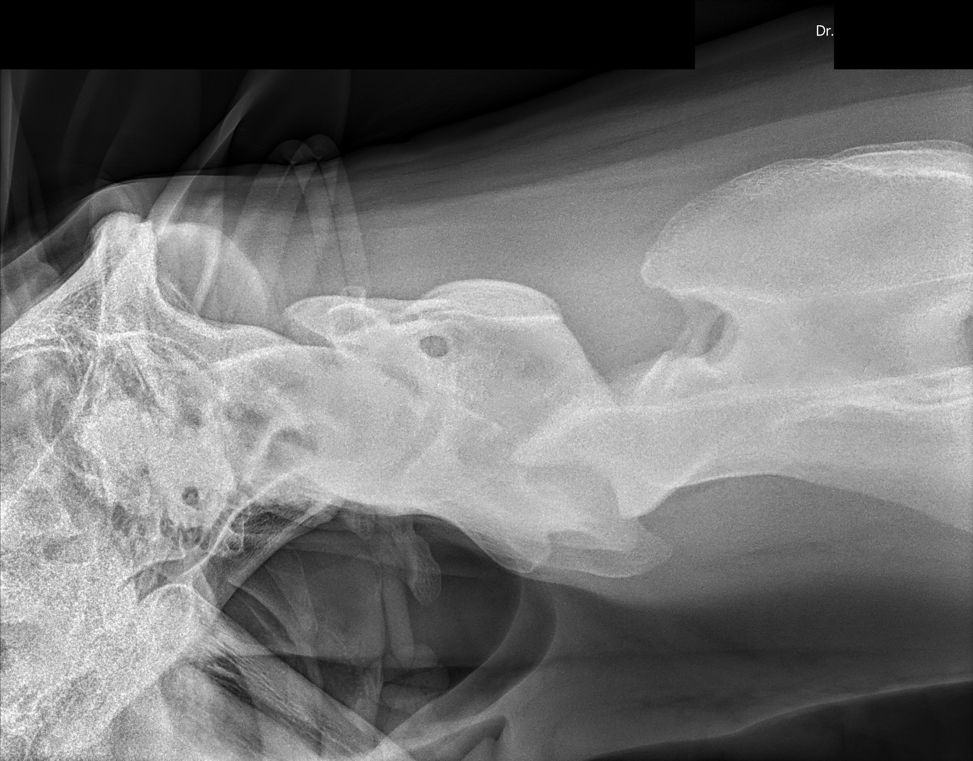
X-ray diagnostic quality assessment
Don't waste time analyzing non-diagnostic X-rays.
If an image should be retaken, Delara flags it early. No more losing time on poor-quality radiographs. No more confusing artifacts with lesions.
Quality Metrics
Instant quality scoring per view including positioning, exposure, and motion detection.
- Positioning accuracy98%
- Exposure optimization95%
- Motion detection92%
Anomaly Detection
We bring expert-level radiology support to every vet, everywhere. Your second opinion assistant. AI assists, vet decides.
Instant Detection
AI identifies abnormalities in seconds with high accuracy
Quality Assurance
Verify findings with confidence scoring
- LowSpur - LH FetlockConfidence: 78% |
- MediumFragment - RF FetlockConfidence: 87% |
- HighBone cyst - RH FetlockConfidence: 93% |
Automated anatomic measurements
Upload a radiograph. Delara instantly computes every measurement without a single click. What used to take minutes of careful manual tracing now happens in seconds, with consistent precision across every case.
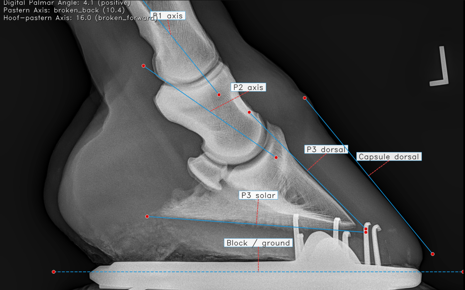
- Digital Palmar Angle4.1°
- Pastern AxisBroken Back
- Hoof-Pastern AxisBroken Forward
Automated Report Writing
AI does the formatting work. You focus on care. Write your report during the consultation — in a few clicks, it's over. No more long nights writing reports from old Word files.
• LF proximal suspensory: mild irregular bone attachment lateral and medial
• Small focal hypoechogenic zone at palmar margin of lateral lobe, no Doppler activity (likely fat)
• Mild thickening suspensory body / cannon medially, mild sensitivity on deep palpation
• Very mild effusion of digital tendon sheath LF, no pain
LF lameness improved ~95% since acute episode. Findings consistent with a mild suspensory sprain (LF) following a stumble at landing. No active tendon lesion identified on ultrasound.
• Continue 1 week of walking, discontinue bute
• Gradual return to work if sound, monitor closely
• Chiro + rapid release: sacrum, L ilium, T14–L6, sternum, withers, neck
• Re-check in 3–4 weeks if any recurrence
Track changes with confidence
Compare radiographs side-by-side to monitor progression over time.
For better care, starting with better radiology
The vet should only focus on diagnosis, not on dealing with what AI can do for you nowadays.
For the veterinarian working alone
An always-on second opinion that brings expert radiology support to every practice, no matter the size.
For better equine welfare
Faster, more accurate diagnoses mean horses get the right care, sooner.
For every vet, everywhere
Equal access to advanced imaging tools, regardless of location or resources.
See Delara in action
Watch a complete workflow from field capture to report — in under 9 minutes.
Get early access — preferred pricing
Lock in our launch price and start free — no credit card required.
Try for free — no account needed
Jump straight in as a guest and explore the platform.
Early Access
Get full access to Delara Vision while we build the future of veterinary radiology.
1 month free — no credit card required, cancel anytime
- AI Anomaly Detection (BETA)
- Report Writing
- Anatomic Measurements
- Compare Over Time
- Unlimited access
- Early access to upcoming features (Quality Diagnostic Check, PACS Connectors, etc.)
Sign up before end of May and lock in your early adopter price.
Start using Delara Vision for free
Join 10+ clinics already using Delara to streamline their radiology workflow. No credit card required.
Free Trial
Start with our free tier and upgrade anytime.
Secure & Private
Enterprise-grade security and data protection.
Easy Setup
Get started in minutes — no complex installation.